Beranda
/ Anatomy Of Chest : Stockfoto Anatomy Chest Pain Pain In The Chest Or The / However, the classical anatomical descriptions in textbooks make it difficult to gain full mastery of this subject, because the books usually deal with its elements separately.
Anatomy Of Chest : Stockfoto Anatomy Chest Pain Pain In The Chest Or The / However, the classical anatomical descriptions in textbooks make it difficult to gain full mastery of this subject, because the books usually deal with its elements separately.
Insurance Gas/Electricity Loans Mortgage Attorney Lawyer Donate Conference Call Degree Credit Treatment Software Classes Recovery Trading Rehab Hosting Transfer Cord Blood Claim compensation mesothelioma mesothelioma attorney Houston car accident lawyer moreno valley can you sue a doctor for wrong diagnosis doctorate in security top online doctoral programs in business educational leadership doctoral programs online car accident doctor atlanta car accident doctor atlanta accident attorney rancho Cucamonga truck accident attorney san Antonio ONLINE BUSINESS DEGREE PROGRAMS ACCREDITED online accredited psychology degree masters degree in human resources online public administration masters degree online bitcoin merchant account bitcoin merchant services compare car insurance auto insurance troy mi seo explanation digital marketing degree floridaseo company fitness showrooms stamfordct how to work more efficiently seowordpress tips meaning of seo what is an seo what does an seo do what seo stands for best seotips google seo advice seo steps, The secure cloud-based platform for smart service delivery. Safelink is used by legal, professional and financial services to protect sensitive information, accelerate business processes and increase productivity. Use Safelink to collaborate securely with clients, colleagues and external parties. Safelink has a menu of workspace types with advanced features for dispute resolution, running deals and customised client portal creation. All data is encrypted (at rest and in transit and you retain your own encryption keys. Our titan security framework ensures your data is secure and you even have the option to choose your own data location from Channel Islands, London (UK), Dublin (EU), Australia.
Anatomy Of Chest : Stockfoto Anatomy Chest Pain Pain In The Chest Or The / However, the classical anatomical descriptions in textbooks make it difficult to gain full mastery of this subject, because the books usually deal with its elements separately.. This page provides an overview of the chest muscle group. The chest anatomy includes the pectoralis major, pectoralis minor and the serratus anterior. Thank you for visit anatomynote.com. The myotomes elongate and invade the mesoderm of the wall of the embryonic thoracic and abdominal cavities. Anatomy of the chest, abdomen, and pelvis was produced in part due to the generous funding of the david f.
This page provides an overview of the chest muscle group. About the 6th week, the somites differentiate into the sclerotomes and the dermatomyotomes. An overview of the anatomy visible in a transverse computed axial tomographical image of the thorax (and part of the abdomen) performed with intravenous cont. The chest wall is formed from the sternum anteriorly, 12 pairs of ribs, costal cartilages and intercostal muscles laterally, and the thoracic vertebrae posteriorly. Thank you for visit anatomynote.com.
Human Chest Anatomy Illustration Stock Image F025 1027 Science Photo Library from media.sciencephoto.com The past several decades have seen a marked improvement in the management and reconstruction of complex chest wall de … The myotomes elongate and invade the mesoderm of the wall of the embryonic thoracic and abdominal cavities. The chest or thorax is the region between the neck and diaphragm that encloses organs, such as the heart, lungs, esophagus, trachea, and thoracic diaphragm. You can click the image to magnify if you cannot see clearly. This page provides an overview of the chest muscle group. The muscles of the chest develop from the somites found in the mesoderm. The circulatory system does most of its work. Thank you for visit anatomynote.com.
The chest wall is formed from the sternum anteriorly, 12 pairs of ribs, costal cartilages and intercostal muscles laterally, and the thoracic vertebrae posteriorly.
This image added by admin. About the 6th week, the somites differentiate into the sclerotomes and the dermatomyotomes. (1) the pectoralis major, and (2) the pectoralis minor. A typical heart is approximately the size of your fist: This page provides an overview of the chest muscle group. The shape of the chest is often regarded as potential insight into a disease process, as in the case of barrel chest and respiratory dysfunction. Thank you for visit anatomynote.com. Definition (nci_cdisc) the anterior side of the thorax from the neck to the abdomen. In insects, crustaceans, and the extinct trilobites, the thorax is one of the three main divisions of the creature's body, each of which is in turn composed of multiple segments. Here, we break down the anatomy of your chest muscles. The circulatory system does most of its work. This article focuses on the unique structural characteristics in … The myotomes elongate and invade the mesoderm of the wall of the embryonic thoracic and abdominal cavities.
This page provides an overview of the chest muscle group. Thank you for visit anatomynote.com. Organs & structures of the chest heart. As with all parts of the body, the anatomy and physiology of the chest wall are intimately intertwined. The past several decades have seen a marked improvement in the management and reconstruction of complex chest wall de …
Venous System Of The Chest Artwork Stock Image C021 2197 Science Photo Library from media.sciencephoto.com To carry out the unique functions performed by the chest wall, the anatomic structures are formed precisely for maximal efficiency. 12 cm (5 in) in length, 8 cm (3.5 in) wide, and 6 cm (2.5 in) in thickness. The muscles of the chest develop from the somites found in the mesoderm. (1) the pectoralis major, and (2) the pectoralis minor. Organs & structures of the chest heart. Having to do with the chest. A line is drawn from anterior surface of the body of 6th thoracic vertebrae passing through the apex of the heart up to anterior lower most part of diaphragm. First i'll do an intro to the different organs and structures in the chest, and then i'll go over some images showing their locations.
Here, we break down the anatomy of your chest muscles.
The chest wall functions as a protective cage around the vital organs of the body, and significant disruption of its structure can have dire respiratory and circulatory consequences. Learn about each of these muscles, their locations, functional anatomy and exercises for them. Fill out your shirt with a bigger, stronger, more powerful chest. The circulatory system does most of its work. It provides protection to vital organs (eg, heart and major vessels, lungs, liver) and provides stability for movement. The pec major) is the one that commands the most real estate. How to view the anatomical labels. An overview of the anatomy visible in a transverse computed axial tomographical image of the thorax (and part of the abdomen) performed with intravenous cont. In insects, crustaceans, and the extinct trilobites, the thorax is one of the three main divisions of the creature's body, each of which is in turn composed of multiple segments. Strictures, acute syndromes, neoplasms and vascular impressions Chest a man's chest — like the rest of his body — is covered with skin that has two layers. Thank you for visit anatomynote.com. Anatomy of the chest and shoulder, anatomy of the chest organs, anatomy of the chest wall, anatomy of the chest wall and pleura, anatomy of upper chest area, human.
The chest wall is comprised of skin, fat, muscles, and the thoracic skeleton. This atlas is a comprehensive and affordable learning tool for medical students and residents and especially for radiologists and pneumologists. The chest anatomy includes the pectoralis major, pectoralis minor and the serratus anterior. The chest wall is formed from the sternum anteriorly, 12 pairs of ribs, costal cartilages and intercostal muscles laterally, and the thoracic vertebrae posteriorly. The chest wall functions as a protective cage around the vital organs of the body, and significant disruption of its structure can have dire respiratory and circulatory consequences.
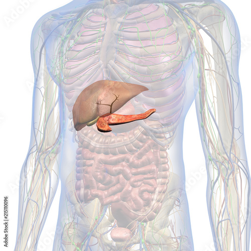
Male Internal Anatomy Of Chest And Abdomen With Pancreas And Liver Highlighted Stock Illustration Adobe Stock from as1.ftcdn.net The chest or thorax is the region between the neck and diaphragm that encloses organs, such as the heart, lungs, esophagus, trachea, and thoracic diaphragm. Anatomy of the chest and shoulder, anatomy of the chest organs, anatomy of the chest wall, anatomy of the chest wall and pleura, anatomy of upper chest area, human. The shape of the chest is often regarded as potential insight into a disease process, as in the case of barrel chest and respiratory dysfunction. The chest wall is comprised of skin, fat, muscles, and the thoracic skeleton. 12 cm (5 in) in length, 8 cm (3.5 in) wide, and 6 cm (2.5 in) in thickness. A line is drawn from anterior surface of the body of 6th thoracic vertebrae passing through the apex of the heart up to anterior lower most part of diaphragm. The muscles of the chest develop from the somites found in the mesoderm. Definition (nci_cdisc) the anterior side of the thorax from the neck to the abdomen.
First i'll do an intro to the different organs and structures in the chest, and then i'll go over some images showing their locations.
Anatomy of right side chest pain. It is made up of the manubrium superiorly, the body and the xiphisternum (figure 1). This atlas is a comprehensive and affordable learning tool for medical students and residents and especially for radiologists and pneumologists. The chest wall is comprised of skin, fat, muscles, and the thoracic skeleton. Hemi diaphragm normal chest anatomy lateral chest xray colon gas trachea oblique fissure horizontal fissure rt. Browse 2,532 female chest anatomy stock photos and images available, or start a new search to explore more stock photos and images. It is enclosed by the ribs, the vertebral column, and the sternum, or breastbone, and is separated from the abdominal cavity (the body's largest hollow space) by a muscular and membranous partition, the diaphragm. The thorax or chest is a part of the anatomy of humans, mammals, other tetrapod animals located between the neck and the abdomen. Radiology basics of chest ct anatomy with annotated coronal images and scrollable axial images to help medical students and junior doctors learning anatomy. Computed tomography (ct) of the chest can detect pathology that may not show up on a conventional chest radiograph (1). Here, we break down the anatomy of your chest muscles. As with all parts of the body, the anatomy and physiology of the chest wall are intimately intertwined. First i'll do an intro to the different organs and structures in the chest, and then i'll go over some images showing their locations.

